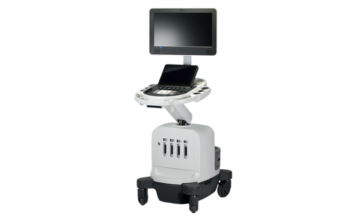
Vizuālā diagnostika Ltd. ensures echocardiography or Doppler echocardiography (EchoCG) examinations by using Philips and GE Healthcare devices, that offer precise diagnostic examinations that enable the patients to receive especially high-resolution images of high quality.
Echocardiography is the method of heart examination and diagnostics that is based on the registration of the ultrasound signals reflected from moving structures of the heart. The examination provides the physician with the opportunity to determine the anatomical structure of the heart, the dimensions of the ventricles and atria, the thickness of the walls of the heart, to evaluate the functional condition of heart valves, as well as to determine the presence of scars or thrombi after myocardial infarction. EchoCG can be used to determine the velocity and direction of blood flow in the heart and to determine deviations from the norm that are dangerous to the patient, but do not cause immediate complaints.
It is important to possess knowledge of the symptoms of cardiovascular diseases and to be able to recognise life threatening situations to understand, when the heart tests need to be performed and when emergency medical care must be immediately called.
- In what situations is the performance of EchoCG examination recommended?
- If the patient has pain in the area of the heart;
- If the patient has elevated blood pressure;
- If the patient complains of arrhythmia;
- If the tones of the heart are altered or cardiac noises are detected;
- If fainting and/or shortages of breath occur;
- If cholesterol levels are elevated;
- If the amount of blood oxygen is decreased or the skin and mucous membranes are bluish in colour;
- If chemotherapy medications or other medications that affect hear function are used;
- If the assessment of heart function indicators after myocarditis and endocarditis (inflammation processes in the heart) is required.
- What should a patient know before the performance of an EchoCG examination?
EchoCG is painless and completely harmless for the health of the patient (no ionising radiation is used), it can be performed in children and pregnant women as well. The patient does not require special preparations. The examination lasts for 15 – 20 minutes.
During the examination, the patient needs to assume supine position on the therapy couch or to lie on their side, remove clothing from the chest, where a gel-coated receiver, which creates mobile images of the heart on the image, is placed.


